Table of Contents
Introduction
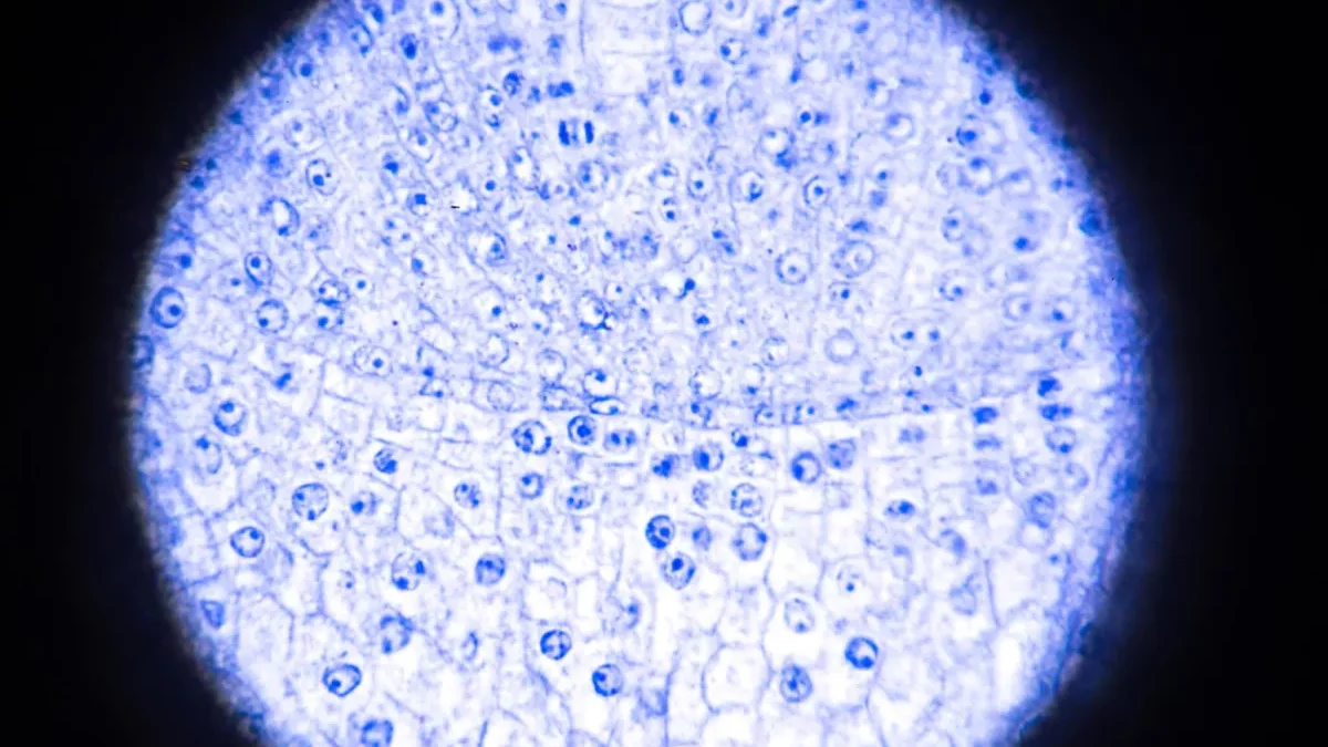
The fundus autofluorescence study is a non-invasive test performed with a short, bright light while the patient is dilated to allow as much light as possible into the eye. FAF is a type of photography that uses a special light to assess highly fluorescent optical structures. This test checks for the presence of fluorophores in the eye. Fluorophores are chemical structures that possess fluorescent properties when bare to the light of a proper wavelength.
The autofluorescence test uses the fluorescent properties of a lipofuscin metabolic indicator to study the retinal epithelium’s health. When other fluorophores in the outer retina can appear with the disease, lipofuscin is the primary source of intrinsic fundus fluorescence. When there is an excessive accumulation of lipofuscin, it can be the origin of several complex and hereditary retinal diseases.
What is Fundus Auto Fluorescence?
The ability of the eye to absorb light of a particular wavelength and subsequently emit it at a higher wavelength (between 500 and 700 nm). This process occurs in the retina, a layer inside the eyeball made up of cells that allow this phenomenon.
The leading autofluorescent component of the retina is lip fusain. Lip fusain is a yellowish pigment that occurs best naturally due to the metabolism and degradation of the photoreceptor cells that make up the retina. The greater or lesser fluorescence of this will depend on the number of flour pores present in the lip fusain.
What is used Fundus Auto Fluorescence?
Commonly, accumulations of lip fusain are generated during the metabolism of the cells in the retina. An excessive collection of lip fusain is usually an indicator of an eye disease that affects the retina, mainly the retinal pigment epithelial layer.
The study of autofluorescence helps us detect, diagnose and monitor specific retina pathologies. It provides information about the state of the cells and pigments of the retina.
How does Fundus Auto Fluorescence Work?
The autofluorescence test is a fast and non-invasive technique for the patient. It bases exclusively on the light-emitting property of the retina. It performs without intravenous contrast, unlike ocular angiography.
During the performance of the test, images of the eye fundus obtain. These images show a pattern of fluorescence distribution on the ocular surface, presenting areas of hypo auto fluorescence (areas with more negligible autofluorescence) or hyper auto fluorescence (areas of excessive fluorescence). The ophthalmologist is in charge of analyzing and interpreting patterns obtain. In general, each print is associated with a possible pathology or specific situation of the state of the retina.
What does Fundus Auto Fluorescence Consist of?
Auto fluorescence takes advantage of the fluorescent properties of a metabolic indicator called lip fusain, which is present in the ocular fundus as a result of light stimulation of the cellular pigments of the retinal pigment epithelium.
Thanks to the development of diagnostic imaging technologies, the study of lip fusain accumulation patterns can perform non-invasively for the patient, visualizing its deposits in vivo since an increase or decrease in certain areas is the reflex of pathological changes.
To reach even the most peripheral areas, wide-field systems are beneficial, which require a single image capture to visualize 200° of the retina (compared to 30-45° of the central field).
In what cases is it used?
Autofluorescence indicates for patients with pathologies of the retina that affect the pigment epithelium, with retinal dystrophies and AMD the main ones, in addition to drug toxicity, and posterior uveitis or some intraocular tumours, among others.
This technique is instrumental in unexplain loss of visual acuity and hereditary retinal dystrophies, such as retinitis pigments or Star grad disease. It has seen that the processes involves are relating to alterations in this layer, which can help identify the specific type of dystrophy and guide genetic counselling.
Method of Fundus Auto Fluorescence
The autofluorescence test uses the fluorescent properties of a metabolic indicator called lip fusain. In to study the health of the retinal epithelium. When other flour pores in the outer retina may appear with the disease, lip fusain is the primary source of intrinsic fluorescence in the fundus. When there is an excessive accumulation of lip fusain, it can the origin of several complex and hereditary retinal diseases.
Fundus autofluorescence can use to assess the presence of:
- Optic nerve druse
- Plaque nil toxicity
- Macular dystrophies
- Retinitis pigments
- White point syndromes.
Conclusion
This procedure bases on a property of the eye fundus: autofluorescence. This quality is an intrinsic capacity. That the average human retina presents and consists of the emission. The light in the spectrum of wavelengths is from 500 to 700 nm. The leading auto fluorescent component of the fundus is rim fusain. Lip fusain is a yellowish-brown pigment that remains from decomposition and absorption. Its damages blood cells. In the fundus of the eye and under normal conditions, it forms part of the retinal pigment epithelium (RPE). Therefore, changes in this pigment epithelium. Such as abnormal lip fusain deposits or a certain level of atrophy, will be evident when the ophthalmologist performs this test.
Also Read: K&G